
ELHGHII – Human/Monkey IgG anti-human
General description
The product binds human IgG and not other human Ig. Immunoglobulins are composed of antigen-binding domains of two fragments (Fab) and one crystallizable fragment (Fc). The gene encoding the IgG gene cluster is found on human chromosome 14. Anti-human IgG antiserum (Fc specific) is produced in goat using purified human IgG, Fc fragment, as an immunogen. Affinity isolated antibody is obtained from goat anti-human IgG antiserum by immunospecific purification that removes essentially all goat serum proteins, including immunoglobulins, that do not specifically bind to the Fc fragment of human IgG.
Specificity
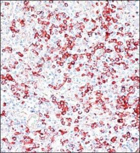
Specificity for the human IgG Fc fragment is determined by ELISA and immunoelectrophoresis (IEP). The antibody preparation is specific for human IgG, Fc fragment when tested against purified human IgA, IgG (Fc and Fab fragments), IgM, Bence Jones kappa, and Bence Jones lambda myeloma proteins. No reactivity is observed with the Fab fragment of human IgG, light chains, IgA or IgM. The affinity-purified anti-human IgG (Fc-specific) reagent offers the advantage of increased sensitivity for human IgG without cross-reactivity with other substances present on the cell membrane or surface.
The lack of cross-species cross-reactivity with mouse or rat serum proteins makes this product excellent for screening human monoclonal antibodies produced by hybridoma cells grown in vivo in mouse or rat ascites fluids. This product has the ability to detect all subclasses of human IgG in human biological fluids or tissues from normal or pathological situations such as cancer or autoimmune diseases. It is effective as a second antibody reagent in immunoassay procedures and can be used as a starting material for conjugates using enzymes or fluorescent dyes.
Immunogen
Anti-human IgG antiserum (Fc specific) is produced in goats using purified human IgG, Fc fragment, as an immunogen
Request
The anti-human IgG antibody (specific for Fc) produced in goats has been used:
- in double capture ELISA to measure antiglobulin responses in the serum of transplant patients treated with monoclonal antibodies CD52 (CAMPATH-1G)
- in the detection of IgG levels in patients with rheumatoid arthritis
- in small bowel biopsies
- in patients with acute myocardial infarction by immunoblotting
Physical form
The solution in 0.01 M phosphate-buffered saline, pH 7.4, containing 15 mM sodium azide
Storage and stability
For continuous use, store at 2-8 ° C for up to one month. For long-term storage, the solution can be frozen in working aliquots. Repeated freezing and thawing, or storing in “frost-free” freezers is not recommended. If slight cloudiness occurs after prolonged storage, rinse the solution by centrifugation before use.
Disclaimer
Unless otherwise stated in our catalog or other company documentation accompanying the product (s), our products are designed for research use only and are not to be used for any other purpose, including but not limited to commercial uses. unauthorized, in vitro diagnostic uses, ex vivo or in vivo therapeutic uses or any type of consumption or application to humans or animals.
Biochemical / Physiological Actions
The IgG antibody subtype is the most abundant of the serum immunoglobulins of the immune system. It is secreted by B cells and is found in blood and extracellular fluids and provides protection against infections caused by bacteria, fungi, and viruses. Maternal IgG is transferred to the fetus through the placenta, which is vital for the newborn’s immune defense against infection. Mutations in the Fc region of IgG are implicated in autoimmune diseases such as rheumatoid arthritis. Modified Fc proteins are of therapeutic importance for the treatment of autoimmune diseases.